In the realm of modern medicine, the advent of imaging technology has been nothing short of revolutionary. From diagnosing ailments to guiding intricate surgeries, imaging has become an indispensable tool for healthcare professionals worldwide. Among the myriad techniques available, one method stands out for its precision and versatility: imaging with slices. This cutting-edge approach has transformed the way we perceive and understand the human body, offering unprecedented insights into its inner workings.
Imaging tech with slices, also known as tomographic imaging, involves capturing a series of cross-sectional images or “slices” of an object or body part. These slices are then reconstructed to generate a three-dimensional representation, providing a comprehensive view of the subject under examination. This technique finds application in various fields, including medicine, biology, archaeology, and materials science, offering invaluable benefits in each domain.
Revolutionizing Medical Imaging
In the realm of medicine, tomographic imaging has become indispensable for diagnosing a wide range of conditions, from bone fractures to neurological disorders. One of the most common modalities used in medical imaging is computed tomography (CT), which utilizes X-rays to create detailed cross-sectional images of the body. CT scans provide clinicians with vital information about the size, shape, and density of internal structures, enabling them to identify abnormalities with unparalleled accuracy.
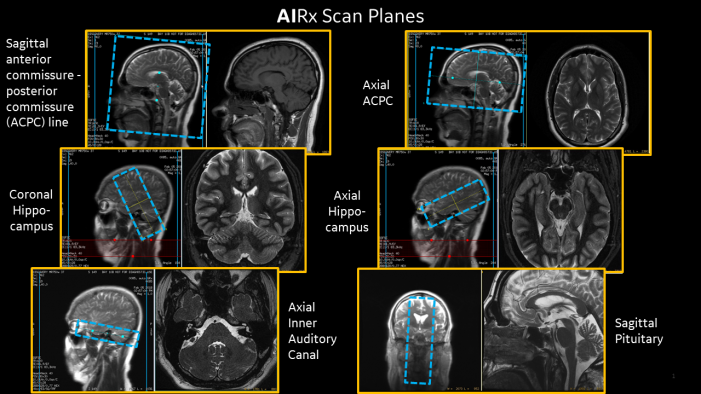
Another widely employed technique is magnetic resonance imaging (MRI), which utilizes powerful magnets and radio waves to generate detailed images of soft tissues, organs, and joints. By acquiring slices from different orientations, MRI produces high-resolution three-dimensional reconstructions, allowing for precise localization of pathology and optimal treatment planning. MRI is particularly valuable in neuroimaging, offering unparalleled insights into the brain’s structure and function.
In recent years, advancements in imaging technology have led to the development of novel modalities such as diffusion tensor imaging (DTI) and functional MRI (fMRI). DTI enables the visualization of white matter tracts in the brain, facilitating the study of neural connectivity and organization. On the other hand, fMRI measures changes in blood flow to detect brain activity, opening new avenues for understanding cognitive processes and neurological disorders.
Beyond the realm of clinical medicine, imaging with slices has found applications in fields such as archaeology and paleontology, where it aids in the non-destructive analysis of artifacts and fossils. By acquiring sequential slices of archaeological specimens, researchers can unveil hidden details, decipher inscriptions, and reconstruct ancient artifacts with remarkable precision. Similarly, in paleontology, tomographic imaging allows scientists to study fossilized remains without damaging or altering their delicate structures, shedding light on evolutionary processes and ancient ecosystems.
Exploring the Marvels
Moreover, imaging technology with slices has revolutionized the field of materials science, enabling researchers to analyze the internal structure and composition of various materials with unprecedented clarity. Techniques such as X-ray microtomography and focused ion beam scanning electron microscopy (FIB-SEM) provide insights into the microscopic features of materials, offering valuable data for optimizing manufacturing processes, developing new materials, and understanding failure mechanisms.
In addition to its diagnostic and research applications, imaging with slices plays a crucial role in guiding minimally invasive procedures and surgical interventions. Techniques such as fluoroscopy, ultrasound, and endoscopic imaging utilize real-time slice acquisition to navigate instruments within the body, ensuring precise placement and minimizing the risk of complications. From cardiac catheterization to laparoscopic surgery, tomographic imaging enhances the safety and efficacy of minimally invasive techniques, ultimately improving patient outcomes.
Despite its myriad benefits, imaging with slices is not without challenges. One of the primary considerations is radiation exposure, particularly in the case of CT scans, which utilize ionizing radiation. While advances in technology have led to dose reduction strategies, minimizing radiation exposure remains a priority in medical imaging. Additionally, the acquisition and processing of volumetric data require substantial computational resources and expertise, limiting its accessibility in certain settings.
Looking ahead, the future of imaging technology with slices holds immense promise, driven by ongoing innovations in hardware, software, and image processing algorithms. Emerging techniques such as spectral CT, photon-counting detectors, and artificial intelligence-based reconstruction algorithms are poised to further enhance imaging quality, speed, and diagnostic accuracy. Moreover, interdisciplinary collaborations between engineers, physicists, and clinicians are paving the way for novel applications and breakthroughs in tomographic imaging.
Conclusion
Imaging technology with slices represents a paradigm shift in our approach to visualizing and understanding the world around us. From unraveling the mysteries of the human body to unlocking the secrets of ancient civilizations, tomographic imaging has transcended boundaries and revolutionized countless fields. As technology continues to evolve and capabilities expand, the impact of imaging with slices on healthcare, science, and society at large is bound to grow, heralding a future where precision, clarity, and insight converge to shape a healthier and more enlightened world.